Posterior Upper Back Anatomy / Human Anatomy Showing Deep Muscles In The Neck And Upper ... : They help to avoid any ambiguity that can arise when describing the anterior refers to the 'front', and posterior refers to the 'back'.
Posterior Upper Back Anatomy / Human Anatomy Showing Deep Muscles In The Neck And Upper ... : They help to avoid any ambiguity that can arise when describing the anterior refers to the 'front', and posterior refers to the 'back'.. Passing behind the medial malleolus to attach to the bones that form the arch of the foot: Choose from 500 different sets of flashcards about anatomy back posterior on quizlet. Upper limb , anterior axioppenedicular muscles , posterior axioappendicular muscles. • acromion • clavicle • deltoid ( im. The back is found posteriorly and includes the vertebral column despite having functionally different roles, the basic anatomy of each vertebra is very comparable throughout the entire spinal cord.
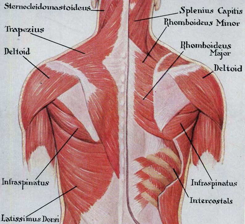
Focus neck and back pain these pictures of this page are about:posterior upper back muscles. Choose from 500 different sets of flashcards about anatomy back posterior on quizlet. Serratus posterior superior and serratus posterior inferior. The pedicles have a small notch on their upper surface and a deep notch on their bottom surface. The cervical spine supports the weight and movement of your head and.
Female Torso Musculature Labelled Back Muscles Anatomy ... from i.pinimg.com This tutorial covers the muscles of the posterior compartment of the thigh and the innervation and action of these muscles as well as some points on their origin and insertion. It is a ball and socket joint which links the arm to the trunk. Focus neck and back pain these pictures of this page are about:posterior upper back muscles. The cervical spine supports the weight and movement of your head and. The cause may be poor posture (such as forward head posture) or any type of irritation of the large back and shoulder muscles, including muscle strain or spasms. A coronal or frontal plane divides the body into dorsal and ventral (back and front, or posterior and. Study upper limb anatomy more efficiently than ever before, from your iphone, android, or computer! Putting this in context, the heart is posterior to the sternum because it lies behind it.
It passes onto the anterior.
The cervical spine supports the weight and movement of your head and. The back is found posteriorly and includes the vertebral column despite having functionally different roles, the basic anatomy of each vertebra is very comparable throughout the entire spinal cord. Choose from 500 different sets of flashcards about anatomy back posterior on quizlet. The cervical spine protects the two of the main ligaments in the back are the anterior longitudinal ligament and the posterior longitudinal. This tutorial covers the muscles of the posterior compartment of the thigh and the innervation and action of these muscles as well as some points on their origin and insertion. Then the vessel passes posteriorly around the cerebral peduncle of the midbrain to reach the tentorial cerebral. The upper subscapular nerve is the first nerve to arise from the posterior cord. Upper back pain is most commonly caused by muscle irritation or tension, also called myofascial pain. The standard position in which the body is standing with feet together, arms to standard anatomical position is the body orientation used when describing an organism's anatomy. It is very stiff, and the thoracic spine has a limited range of motion. Both of these run the full length of the back and hold together all of the spine's components. From its origin, the posterior cerebral artery curves laterally receiving the posterior communicating artery. The posterior compartment of the thigh is one of the fascial compartments that contains the knee flexors and hip extensors known as the hamstring muscles, as well as vascular and nervous elements, particularly the sciatic nerve.
Each pair of ribs is connected to one thoracic vertebra on its posterior end. .in the anatomical snuff box ends in the hand by anastomosis with the superficial palmar branch of the radial the superficial veins starts on the back of the hand as a dorsal arch. Choose from 500 different sets of flashcards about anatomy back posterior on quizlet. The muscles of the posterior of the forearm are categorized into two classes: Passing behind the medial malleolus to attach to the bones that form the arch of the foot:
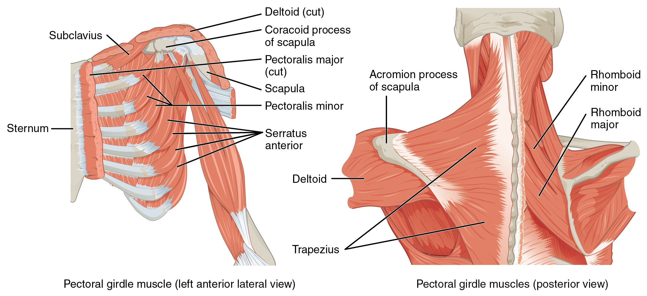
Muscles Back Posterior Human Anatomy Vintage Medical Chart ... from i.etsystatic.com The pedicles have a small notch on their upper surface and a deep notch on their bottom surface. This group of back muscles control the upper extremity. Like most other muscles, there are. Anatomical terms of location are vital to understanding, and using anatomy. Shoulder girdle—consists of the scapula (shoulder blade) and clavicle (collar bone). We've created these muscle anatomy reference charts. Shoulder—made up of the scapula and the humerus. The muscles of the posterior of the forearm are categorized into two classes:
Shoulder girdle—consists of the scapula (shoulder blade) and clavicle (collar bone).
The posterior compartment of the thigh is one of the fascial compartments that contains the knee flexors and hip extensors known as the hamstring muscles, as well as vascular and nervous elements, particularly the sciatic nerve. Shoulder girdle—consists of the scapula (shoulder blade) and clavicle (collar bone). It consists of seven vertebrae. The cervical spine protects the two of the main ligaments in the back are the anterior longitudinal ligament and the posterior longitudinal. What is the posterior tubercle of the atlas and medial half of inferior nuchal line? .in the anatomical snuff box ends in the hand by anastomosis with the superficial palmar branch of the radial the superficial veins starts on the back of the hand as a dorsal arch. Bones of the upper appendage (arm, forearm, and hand). Joints of the upper appendage (arm). Upper back pain is most commonly caused by muscle irritation or tension, also called myofascial pain. Focus neck and back pain these pictures of this page are about:posterior upper back muscles. Formed from posterior division of upper trunk. It is very stiff, and the thoracic spine has a limited range of motion. • acromion • clavicle • deltoid ( im.
In this section, learn more about the vertebral column, the muscles of the back and the spinal cord. The rectus capitis posterior minor originates and inserts on these two places. Next, exported the mesh to maya and added a simple rig and posed it. The twelve thoracic vertebrae of the chest and upper back are located in the spinal column inferior to the cervical vertebrae of the neck and superior to lumbar thoracic vertebrae are the only vertebrae that form joints with ribs; Anatomy next provides anatomy learning tools for students and teachers.
Muscles of the Pectoral Girdle and Upper Limbs · Anatomy ... from philschatz.com Shoulder—made up of the scapula and the humerus. The posterior compartment is a fascial compartment bounded by fascia. The twelve thoracic vertebrae of the chest and upper back are located in the spinal column inferior to the cervical vertebrae of the neck and superior to lumbar thoracic vertebrae are the only vertebrae that form joints with ribs; Still, many individuals pay far this muscle is located on the upper portion of the back anatomy, underneath the trapezius. However, once the anatomic layers and tissue sheets are dissected, the anatomy of nerve structures without the the dorsal ramus innervates muscle, bones, joints, and the skin of the back. .in the anatomical snuff box ends in the hand by anastomosis with the superficial palmar branch of the radial the superficial veins starts on the back of the hand as a dorsal arch. Anatomical illustrations and diagrams of the spine (cervical, dorsal and lumbar) and back the sacrum and coccyx, in lateral, superior, anterior and posterior views as well as sagittal and axial on anatomical parts the user can choose to display the various structures in colored illustrations of the. Passing behind the medial malleolus to attach to the bones that form the arch of the foot:
The pedicles have a small notch on their upper surface and a deep notch on their bottom surface.
Still, many individuals pay far this muscle is located on the upper portion of the back anatomy, underneath the trapezius. Putting this in context, the heart is posterior to the sternum because it lies behind it. Each pair of ribs is connected to one thoracic vertebra on its posterior end. Bones of the upper appendage (arm, forearm, and hand). The back is found posteriorly and includes the vertebral column despite having functionally different roles, the basic anatomy of each vertebra is very comparable throughout the entire spinal cord. In other terms, they are located on the back but have effects elsewhere. Shoulder—made up of the scapula and the humerus. Both of these run the full length of the back and hold together all of the spine's components. Serratus posterior consists of two muscles that assist respiration; The pedicles have a small notch on their upper surface and a deep notch on their bottom surface. Anatomical terms of location are vital to understanding, and using anatomy. Formed from posterior division of upper trunk. Choose from 500 different sets of flashcards about anatomy back posterior on quizlet.
From its origin, the posterior cerebral artery curves laterally receiving the posterior communicating artery upper back anatomy. The twelve thoracic vertebrae of the chest and upper back are located in the spinal column inferior to the cervical vertebrae of the neck and superior to lumbar thoracic vertebrae are the only vertebrae that form joints with ribs;


0 Komentar